Human Heart Anatomy Illustrations and Animations
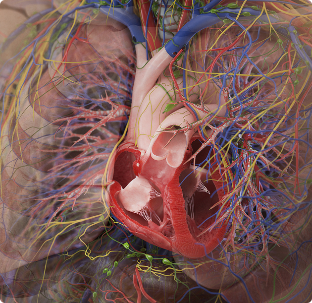
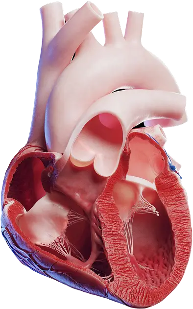
The human heart category contains detailed medical illustrations of cardiac anatomy, covering the four chambers (right and left atria, right and left ventricles), the four heart valves, and the great vessels. Individual assets isolate the aorta, pulmonary trunk and pulmonary arteries, the superior and inferior vena cava, and the pulmonary veins, along with the aortic, mitral, tricuspid, and pulmonary valves.
Anterior, posterior, lateral, and inferior views are available alongside sagittal sections that expose the internal morphology of the atria, ventricles, papillary muscles, and chordae tendineae. Surface renderings show the coronary arteries and cardiac veins across the epicardium. Every structure is rendered from one labeled 3D cardiac model, so anatomy stays consistent across views and cross-sections. Each asset follows Terminologia Anatomica and is reviewed by our Medical Advisory Board.
The collection supports cardiology education, patient communication, pre-operative planning visuals, pharmaceutical and medical device content, and editorial use in textbooks and journals. Assets are available as high-resolution illustrations and 3D animations, licensed under Educational, Standard, or Extended terms through a subscription with credits or as individual purchases.